

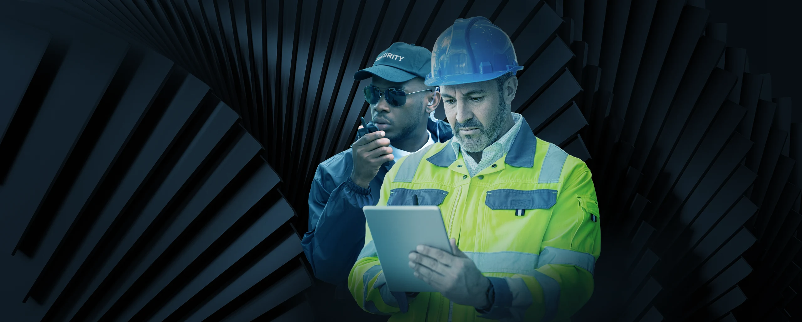
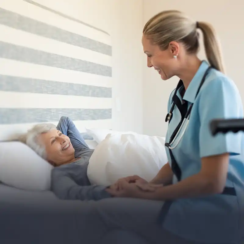
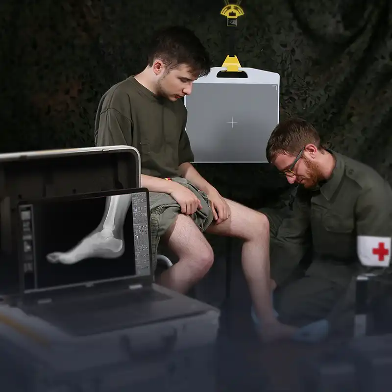
Empowering Frontline Service
Heroes Wherever They Are
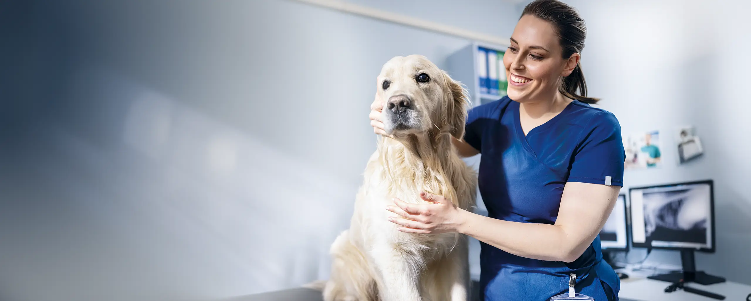
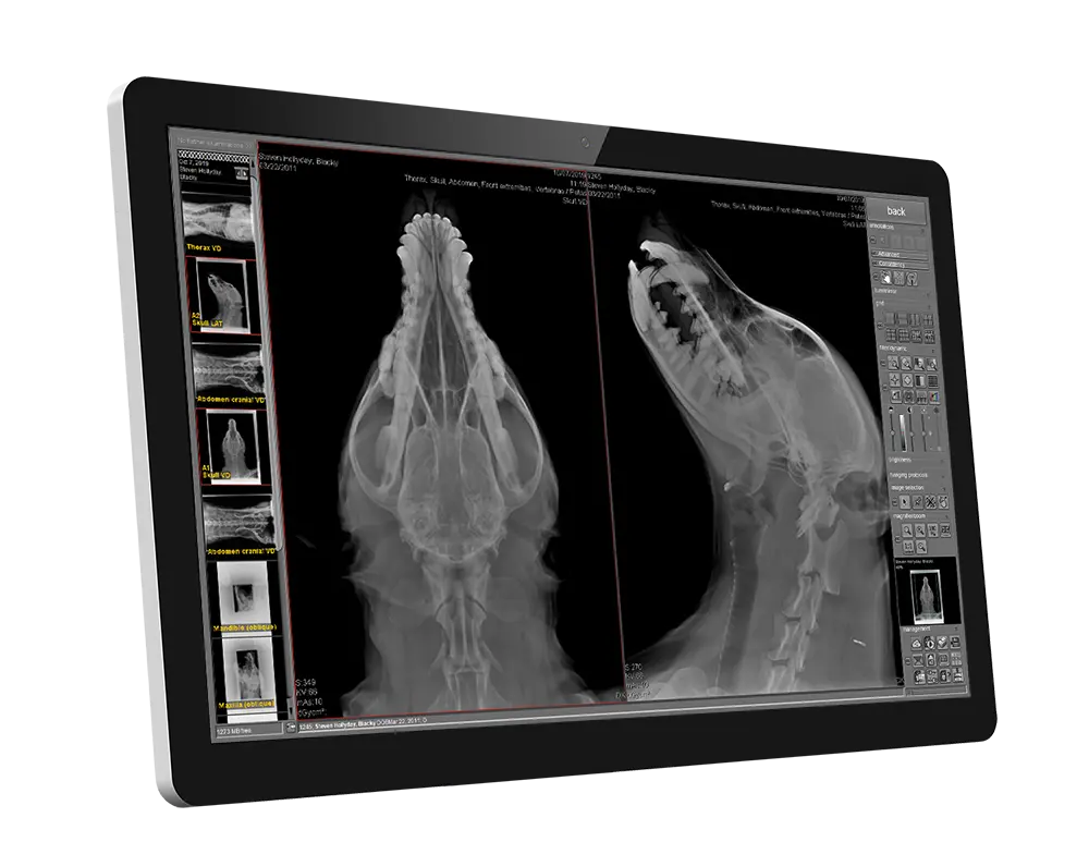
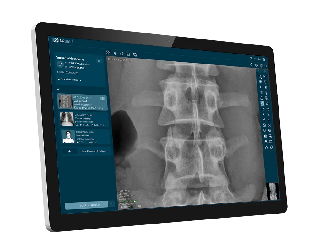
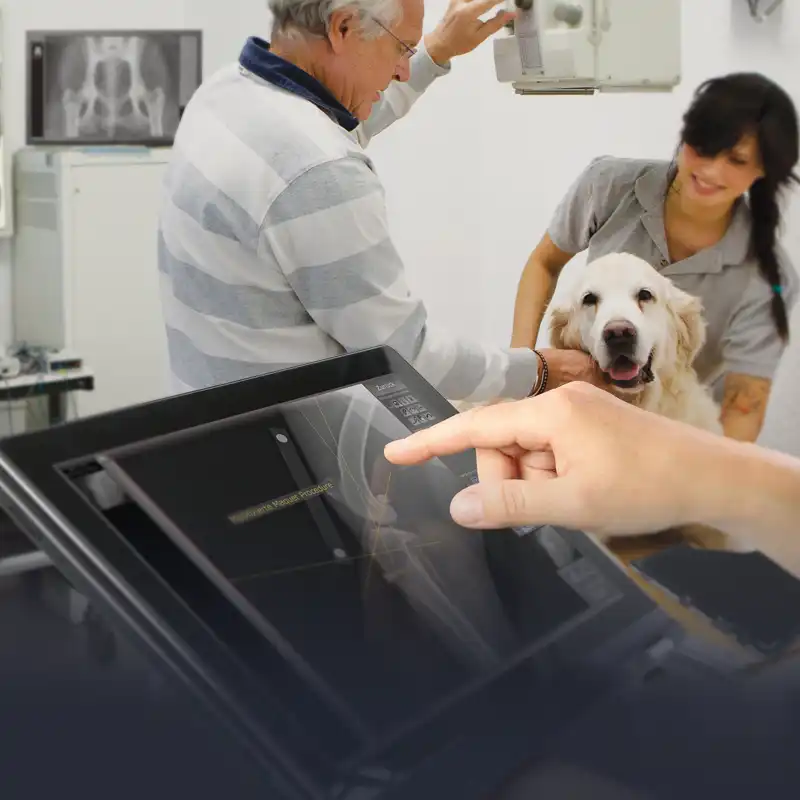
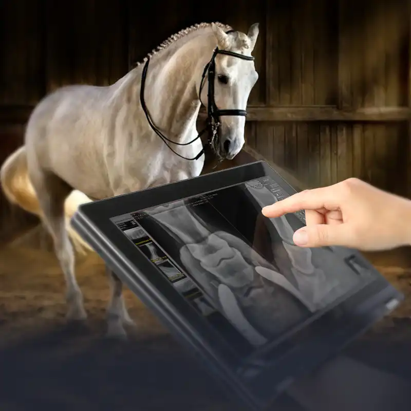
Digital X-ray systems | ultrasound | CT Precise findings & diagnosis – because every animal’s life counts.
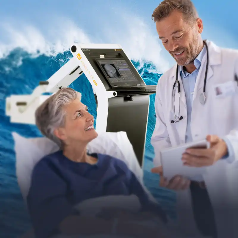
Digital radiography redefined. Laboratory quality for mobile use — ultra-light, high-resolution.
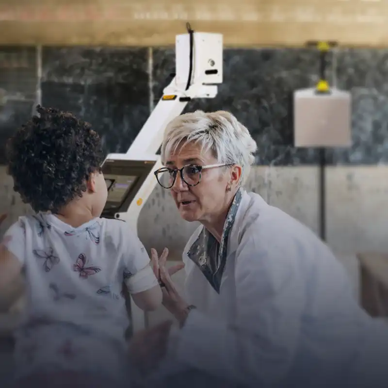
Empowering Frontline Service
Heroes Wherever They Are
OR Technology:
Intelligent imaging for precise diagnoses
OR-Technology stands for innovation, precision and passion. We offer holistic solutions for medical and industrial imaging – from X-ray, computer tomography and ultrasound to high-performance software solutions such as our own PACS.
Our technologies are more than just tools – they are reliable partners for fast and precise diagnostics. With Instant Diagnostic Imaging, we enable professionals worldwide to make immediate and informed decisions.
Experience how state-of-the-art imaging sets new standards. Welcome to OR Technology.
Experience TechnologyThat speaks in favour of your purchase from OR Technology:
For over three decades, OR Technology has been shaping the world of digital X-ray technology – with passion, a spirit of innovation and a firm determination to take medicine and industry to a new level. Founded in 1991.

Impressions that count – your opinion, our success
For over three decades, OR Technology has been shaping the world of digital X-ray technology – with passion, a spirit of innovation and the determination to take medicine and industry to a new level.

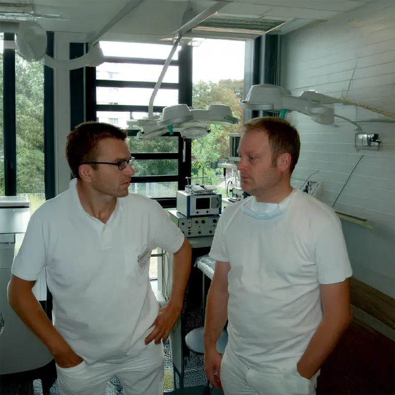
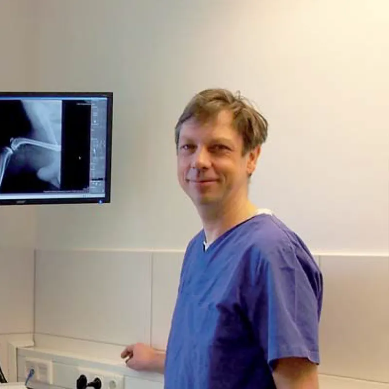
Dr. Torsten Jäschke
Orthopaedic practice, Bad Doberan
Living in Rostock, we have known of OR Technology for a while. We also received some information on their product range through the orthopaedic and emergency surgeons’ association BVOU. The decision to upgrade our existing stationary X-ray unit with a wireless X-ray detector was a quick one. Thanks to OR Technology’s professional advice the installation and introduction of the unit in the practice went smoothly.
Mary Jones
Mobile Medical Diagnostics, Dublin
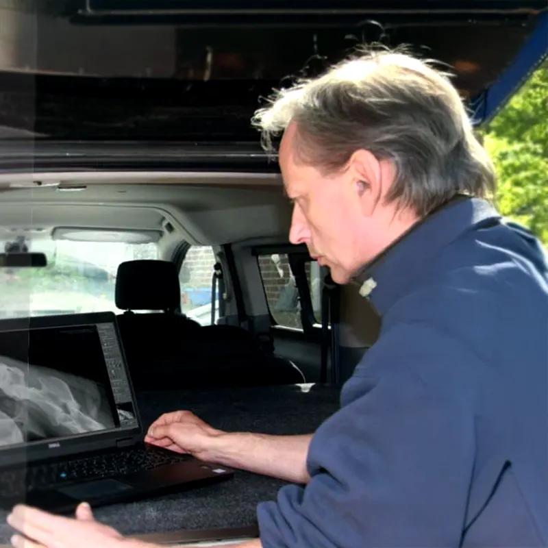
“I found it very difficult to source a mobile X-Ray machine as none of machines I saw in America had CE mark, there seemed to be very limited choice available to me. I wondered what vets were using as they were hardly transporting animals to a fixed service. When I googled veterinary X-Ray systems there you were! Via the OR Technology website I discovered the mobile X ray machine Amadeo M mini.
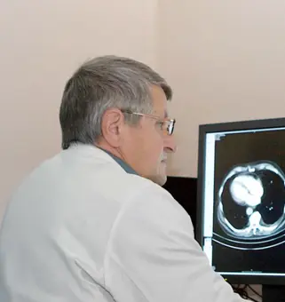
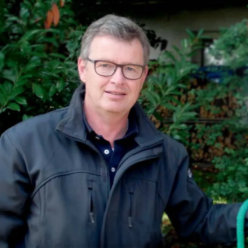
Bernhard Hering
Clinic for Diagnostic and Interventional Radiology, Fulda
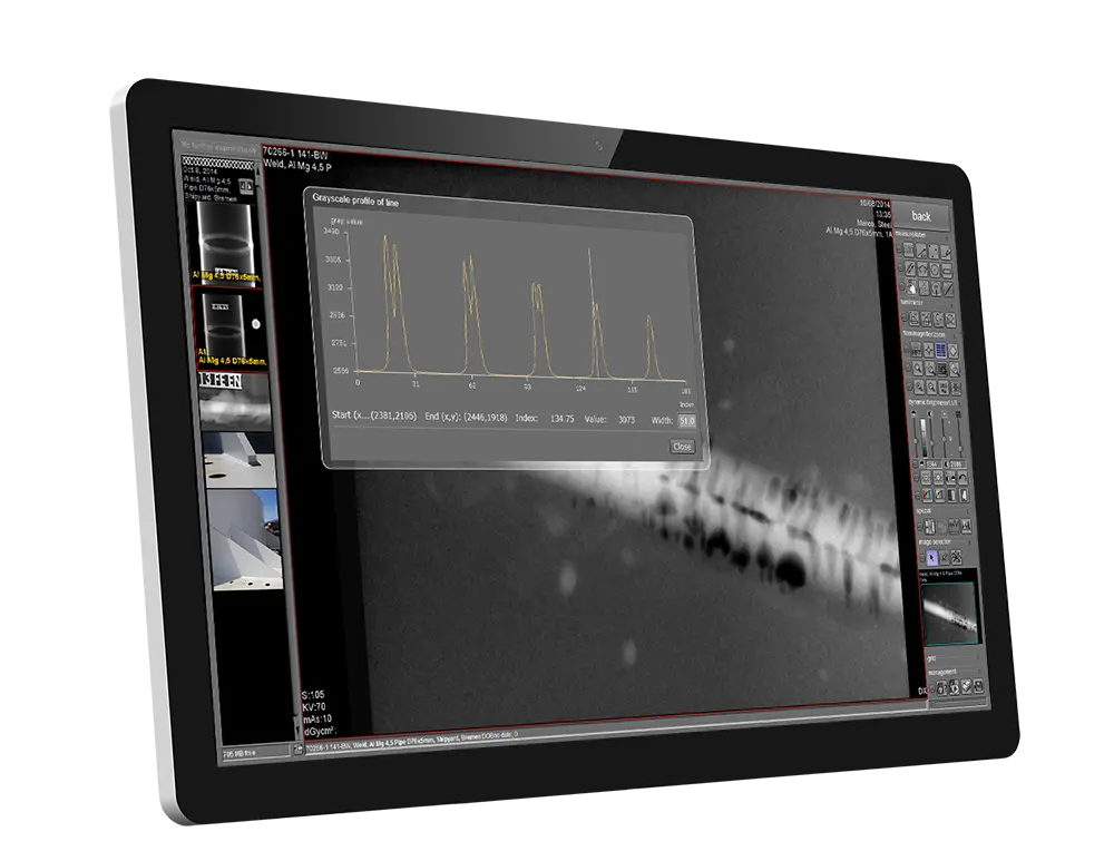
“The great advantage of the retrofit sets is the possibility of bringing analogue X-ray equipment into the digital X-ray age far more effectively than with imaging plates,” explains Bernhard Hering, medical physic expert (MPE).
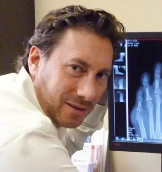
Dr. Ziv Feldman
Feldman Foot & Ankle Clinic in Calgary, Alberta Canada
The advantages provided by the image management solution software for podiatric X-ray diagnosis is almost revolutionary. Angle measurements and special features including zoom and magnification can be done in a few seconds right in front of the patient.
Dr. Igors Malikovs und Dr. Ivars Zvidris
Latvian hospital Jekabpils
In addition, we save costs since we don’t have to print out images any more. Our printing volume now amounts to only 10 % of what it was previously.
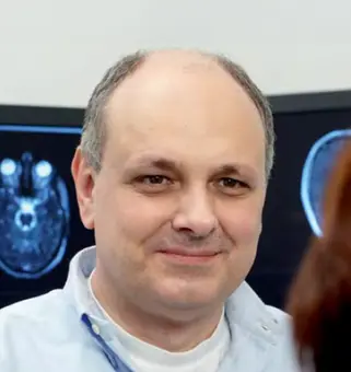
Dr. A. Dawid, Dr. M. Thomson, R. Wiehn
Practice for Radiology and Nuclear Medicine, Zweibrücken
In comparison to our previous system, dicomPACS® is much clearer and much more flexible. It is easy to operate and more customizable. DICOM viewers are now installed on non-diagnostic workstations and allow us to prepare for examinations faster and with better quality. A significant advantage is the much faster and better service …
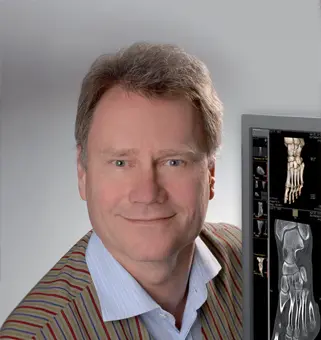
Dr. med. Andreas Bollkämper
Röntgeninstitut Schlossgarten, Hamburg, Germany
In the company OR Technology we have now found a partner for digital image management whose dicomPACS® software solution meets all our demands of up to date, cost efficient operation, providing constant and excellent image quality.
Thomas Blum, the clinic’s Managing Director
Warnow-Klinik Bützow gGmbH, Bützow
The digital X-ray solution by OR Technology allows us to provide optimal service to our patients. This allows our doctors, for instance, to access the required images at the push of a button, to overlay and compare them and thus monitor the course of the healing process.
Dr. med. Stephan Grunert
Orthopaedic practice, Eichstädt
Our orthopaedic practice is highly satisfied with the implementation and and has come to appreciate the enormous benefits of digital X-ray and the fantastic voice recognition facility.
Dr. Bernhard Klein
Orthopaedic group practice, Munich
The entire installation including training took just 2½ days and was done during normal working hours. Since its introduction in 1998 the system has operated without fault or interruption.
Mag. med. vet. Manuel M. Kammermaier
Veterinary practice Kammermaier, Labertal
A complete X-ray system that is easy to handle and equipped with good software to produce high-resolution images. This is how I discovered the complete solution Amadeo V-DR mini II.

Dr. med. vet. Valeska Furck
Small Animal Medicine Competence Centre, Hamburg
Very good image quality and easy handling of the X-ray system were important to me. In addition, a straightforward connection to our practice management software Vetera should be possible, and the PACS system should also be usable for other imaging modalities, particularly computed tomography and ultrasound.
Markus Wirth
Large and Small Animal Practice, Achterwehr
The DR system impresses with fast, mobile deployments, excellent image quality and an optimized workflow – compact, practical and always ready for use.
Dr. Stefan von Bieberstein
Bieberstein Equine Clinic, Cham
The portable solution offers everything my colleagues and I need for an error-free and fast workflow. Thanks to the wireless detector, there are no more annoying cables, and the case is lightweight and easy to handle.
Dr. Hubertus Lutz
Equine Appraiser, Munich
The portable digital X-ray solution Leonardo DR mini from OR Technology has become an indispensable companion in my daily work.
Dr. Alexander Pack and Dr. Karl Scherer
Tierklinik Elversberg
The intuitive operation via the touchscreen monitor convinced us from the very beginning. We can control the entire X-ray system through it. The functions are clearly structured and easy to use. The simple distribution of X-ray images via email to colleagues was another important criterion for us. Last but not least, the excellent price-performance ratio of the system convinced us immediately.
Dr. med. vet. Tido Winkler
Veterinary Group Practice Winkler / Richter
In addition to the significant improvement in workflow and the high-quality X-ray images, what convinced us most was the system’s compatibility with our practice management software.
Caressa Dierenziekenhuis
Netherlands
From our perspective, the Medici DR system is a professional radiography solution that helps us in our daily work to obtain clear findings quickly for necessary follow-up treatments.
Laboratory for Radiology and Sonography, University of Life Sciences in Lublin
Poland
Compared to analog technology, the new digital Medici system significantly shortens image acquisition time. We are highly satisfied with the dicomPACS®DX-R acquisition software, which is very intuitive to use. It is extremely simple to plan and perform an X-ray examination.
Dr. Penny Richards
Central Nova Animal Hospital Ltd.
The user-friendliness of the system with its intuitive operation, the very high-quality X-ray images and the compatibility with our practice management software were the main reasons for our purchase. In addition, the TTA measurement function with different template sizes convinced us. This tool greatly helps us in planning our TTA treatments, for which we are very well known in this region.
Prof. Dr.med.vet. Andrea Meyer-Lindenberg
Surgical and Gynecological Small Animal Clinic of Ludwig-Maximilians-University Munich
In addition to the many beneficial functionalities that characterize the image management software, Prof. Dr. Andrea Meyer-Lindenberg cites the always competent and timely support provided by the OR Technology team as another key decision criterion.
Dr. med. vet. Thomas Weinberger
Equine Clinic Burg Müggenhausen
We can only repeat that we are very satisfied with the investment and the changeover. The software offers a reporting module based on Word that allows targeted reporting on individual images and the direct assignment of these reports to the patient. These reports can be customized according to individual requirements. This has significantly improved both internal and external documentation.